

")
")
")
")

A study jointly conducted by top research institutions including Aix Marseille University (France), the University of Bern, and the University of Zurich (Switzerland) successfully screened five nanobodies that specifically bind to canine PD-L1, four of which effectively block the PD-1/PD-L1 interaction. By constructing bispecific, multivalent, and Fc-fused nanobody complexes, researchers significantly enhanced their blocking efficacy and immune activation capabilities. The best constructs demonstrated picomolar-level inhibitory activity in vitro, effectively induced Antibody-Dependent Cell-mediated Cytotoxicity (ADCC) and Interferon-gamma (IFNγ) release, providing strong candidate drugs for canine cancer immunotherapy. This research, titled "Nanobody-based canine PD-L1-targeting immune checkpoint inhibitors for cancer therapy in dogs," was published on September 18, 2025, in the journal Molecular Therapy: Oncology.
Screening and Identification of Anti-cPD-L1 Nanobodies
The research team first immunized an alpaca with the soluble canine PD-L1 ectodomain and then used phage display technology through multiple rounds of panning to successfully isolate five nanobody clones (Nb34, Nb39, Nb40, Nb41, Nb45). Analysis by Bio-Layer Interferometry (BLI) and flow cytometry revealed that these nanobodies could bind both soluble recombinant cPD-L1 and cell membrane-anchored cPD-L1.
In terms of binding affinity, the nanobodies' affinity for recombinant cPD-L1 ranged from 2 μM to 5 nM, and their apparent affinity for cells expressing cPD-L1 ranged from 150 nM to 4 nM, with Nb34 showing the strongest binding efficacy (See Table 1). Competitive ELISA experiments showed that except for Nb41, the other four nanobodies effectively blocked the cPD-1/cPD-L1 interaction, with half-maximal inhibitory concentration (IC₅₀) values between 340 nM and 53 nM. Simultaneously, flow cytometry confirmed that these nanobodies did not cross-react with human PD-L1, ensuring their specificity for canine therapy.
To clarify the epitope distribution of the nanobodies on cPD-L1, the team conducted epitope binning experiments using BLI. Results showed that Nb34 and Nb39 binding sites were close, partially hindering the binding of Nb40 and Nb45, while Nb40 and Nb45 bound to non-overlapping epitopes. Surprisingly, the monoclonal antibody JC071, known to block the cPD-1/cPD-L1 interaction, competed for binding only with Nb41, which itself cannot block the interaction. This suggests that JC071's blocking effect may rely on steric hindrance rather than direct competition for the PD-1 binding epitope.
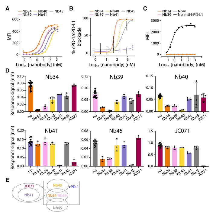
Fig 1:Characterization of anti-cPD-L1 nanobodies
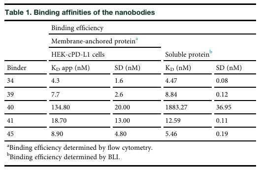
Tab 1:Binding affinities of the nanobodies
Design and Preparation of Multivalent Nanobody Constructs
To enhance the clinical application potential of the nanobodies, the research team designed various multivalent constructs. Nb34 and Nb41, which showed good binding and targeted non-overlapping epitopes, were selected to construct bispecific tandem structures in both orientations (34-41 and 41-34) using flexible linkers. Simultaneously, these nanobodies were fused with different Fc domains, including effector-functional human IgG1 and canine IgGB, as well as effector-silent canine IgGA, to enhance molecular valency, prolong in vivo half-life, and explore Fc-related effector functions.
All nanobody constructs were expressed in mammalian cells, purified using Protein A affinity chromatography (for Fc-bearing constructs) or Immobilized Metal Affinity Chromatography (IMAC, for Fc-less constructs), and quality-controlled by SDS-PAGE and Size Exclusion Chromatography. BLI and flow cytometry assays showed that all engineered molecules effectively bound cPD-L1. Although binding affinity did not significantly improve compared to monovalent nanobodies, the affinity of most constructs for membrane-anchored cPD-L1 increased more than 5-fold, with binding efficiency often reaching the nanomolar range (See Table 2).
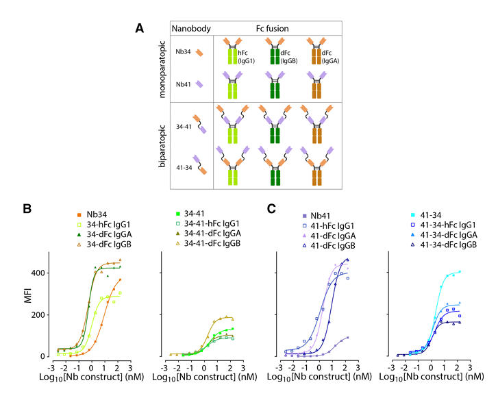
Fig 2:Binding of Nbs and derivative constructs to membrane-anchored cPD-L1
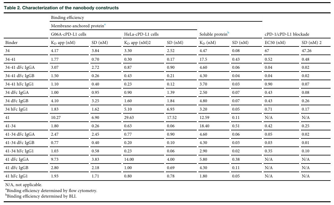
Tab 2:Characterization of the nanobody constructs
Blocking Effect of Nanobody Constructs on the cPD-1/cPD-L1 Signaling Pathway
Using a newly developed cell-based reporter assay system, the research team evaluated the ability of the nanobody constructs to block the cPD-1/cPD-L1 signaling pathway. This system mixes effector cells expressing cPD-1 with artificial antigen-presenting cells (aAPCs) expressing cPD-L1 and detects NFAT promoter-driven luciferase reporter gene signal to reflect pathway blockade.
Results showed that all nanobody constructs significantly increased luciferase activity, except the non-blocking monovalent Nb41, indicating effective blockade of the cPD-1/cPD-L1 signaling pathway. Notably, bispecific constructs containing Nb41 had a better blocking effect than Nb34 alone, while Nb41-Fc and JC071 only slightly increased luciferase activity at high concentrations, further confirming that JC071 likely acts through steric hindrance.
More importantly, the half-maximal effective concentration (EC₅₀) of bivalent constructs was about 100 times lower than that of monovalent nanobodies, reaching the picomolar range. The inhibitory potency of tetravalent dFc molecules (especially those with canine IgGB Fc domain) was about 1000 times higher than monovalent nanobodies and 10 times higher than bivalent constructs. Some tetravalent constructs were about 100 times more effective than atezolizumab (See Table 2), fully demonstrating the superior performance of multivalent nanobody constructs in blocking the cPD-1/cPD-L1 signaling pathway.
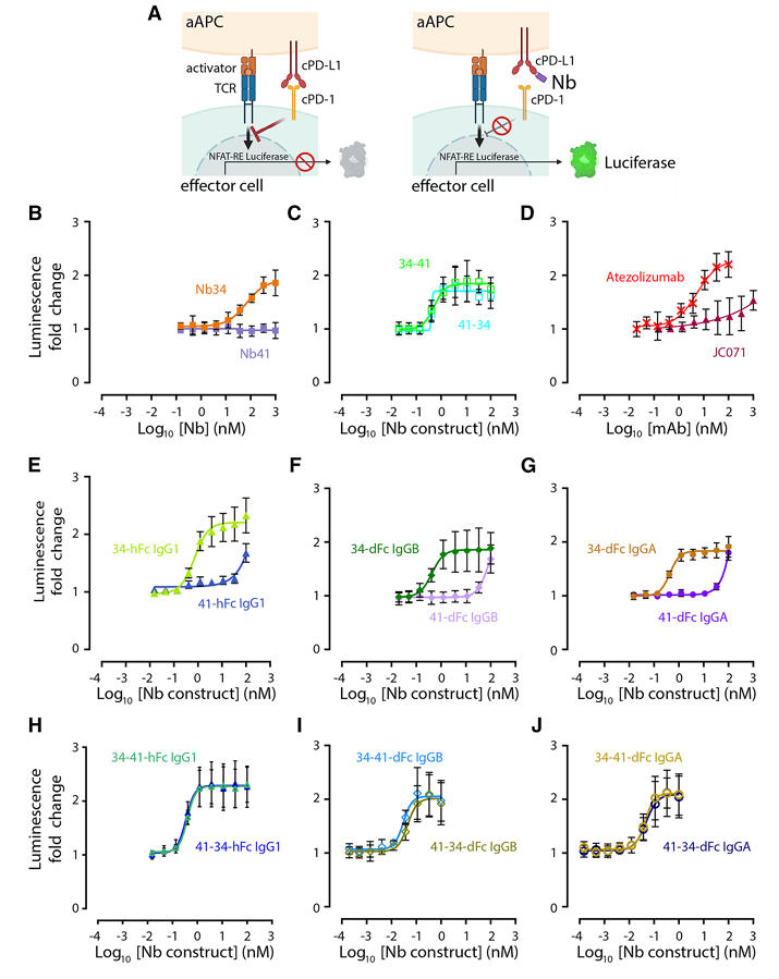
Fig 3:Determination of Nbs and derivative constructs to block the cPD-L1/cPD-1 interaction
Ability of Nanobody Constructs to Induce ADCC Effects
The research team also investigated the ability of nanobody constructs carrying functional Fc domains to induce ADCC effects. Purified human Natural Killer (NK) cells were co-cultured with cPD-L1-expressing HeLa cells in the presence of varying concentrations of nanobody constructs. Results showed that bispecific constructs carrying the human IgG1 Fc domain efficiently induced ADCC at low picomolar concentrations; constructs carrying the canine IgGB Fc domain could also induce a certain degree of ADCC at high concentrations (100 nM).
Under conditions closer to clinical reality, cPD-L1-expressing canine G06A cells were co-cultured with canine Peripheral Blood Mononuclear Cells (PBMCs), or cPD-L1-expressing HeLa cells were co-cultured with human PBMCs. Cell lysis was monitored via Real-Time Cell Analysis (RTCA). Results indicated that nanobody constructs carrying either human IgG1 or canine IgGB Fc domains significantly induced ADCC. Furthermore, in the canine cell system, atezolizumab induced only weak cell death, further validating the advantage of the constructed nanobodies for canine cancer therapy.
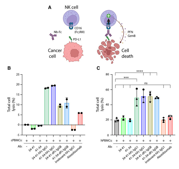
Fig 4:ADCC-triggering by Nb constructs
Effect of Nanobody Constructs on IFNγ Release from Canine T Cells
To evaluate the impact of nanobody constructs on canine T cell activation, the research team used Staphylococcal enterotoxin B (SEB) to stimulate canine PBMCs and detected IFNγ release. SEB can simultaneously bind to MHC class II molecules on antigen-presenting cells and TCR complexes on T cells, activating a large number of T cells and triggering IFNγ release, a process modulated by the cPD-1/cPD-L1 signaling pathway.
Experimental results showed that atezolizumab slightly increased IFNγ production in PBMCs from all donors. In contrast, the two bispecific nanobody constructs carrying the canine Fc-silent isotype (IgGA) showed stronger activity in most donors, with the 34-41-dFc IgGA construct causing a significant increase in IFNγ production. This proves that these nanobody constructs can effectively enhance the immune response of canine T cells, providing further support for their application in canine cancer immunotherapy.
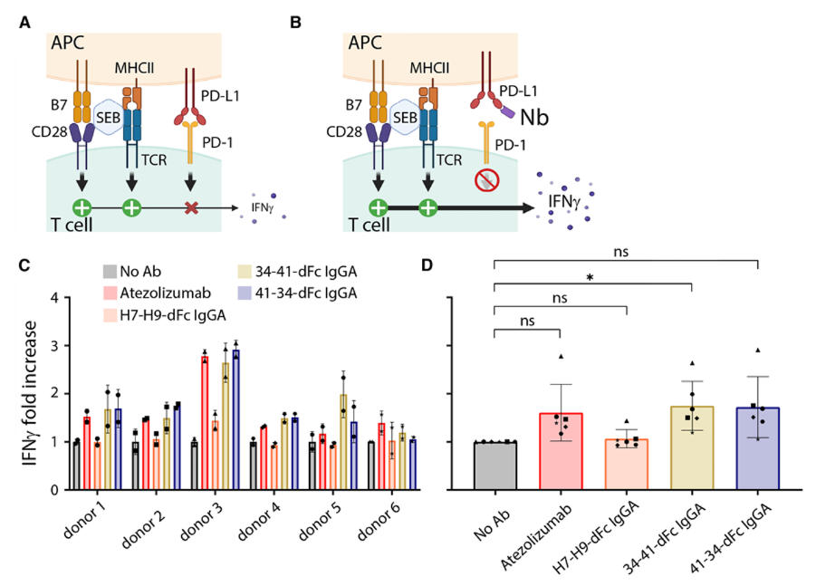
Fig 5:IFNγ production by PBMCs upon treatment with anti-cPD-L1 biparatopic Nb constructs
Research Applications and Industry Value
The nanobodies developed in this study can not only be directly applied to canine cancer treatment, filling a gap in the canine cancer immunotherapy market, but also improve cancer research model development and the study of immunotherapy mechanisms.
Spontaneous canine cancers share high similarities with human cancers in pathological features and immune microenvironment. Moreover, dogs share the same living environment as humans, making their cancer development process better able to mimic human cancer. The nanobodies developed in this study provide ideal tools for investigating the regulatory mechanisms of the cPD-1/cPD-L1 signaling pathway, the principles of immune checkpoint blockade therapy, and the role of ADCC in cancer immunotherapy. Using these tools, researchers can deeply explore the interactions between the immune system and tumors in both dogs and humans, uncover mechanisms of immunotherapy resistance, and provide a theoretical basis for developing novel immunotherapy strategies. They can be used to build more precise canine cancer research models, provide more reliable animal experimental data for human cancer immunotherapy research, and accelerate the development process of human cancer therapeutics.
Wuhan Nano Body Life Science and Technology Co. Ltd. (NBLST) is a nanobody industry platform established under the initiative of the Wuhan Industrial Innovation and Development Research Institute. Its headquarters is located in the main building of the Wuhan Industrial Innovation and Development Research Institute in the East Lake High-tech Development Zone, Wuhan. It boasts a 1400 m² independent laboratory in the Precision Medicine Industrial Base of Wuhan Biolake. Additionally, NBLST has established alpaca experimental and transfer bases in Zuoling, Wuhan, and Tuanfeng, Huanggang, both compliant with laboratory animal standards. These bases currently house over 600 alpacas, providing "zero-immunization-background" guaranteed alpaca immunization services for research institutions and antibody drug development companies.
NBLST focuses on the development, engineering, and application of nanobodies, and is dedicated to building an integrated public experimental service platform for production, education, and research. It possesses a full-chain technology platform encompassing antigen preparation (peptides, proteins, and RNA), antibody discovery and engineering, through to biological function validation/screening. The RNA antigens include RNA structurally and sequentially optimized for alpacas. Antibody discovery and engineering services employ multiple technological routes, including phage display, RNA, and mammalian cell display. Through cross-complementation of multiple platforms, it provides flexible antibody discovery and engineering services for pharmaceutical companies and research institutes, facilitating the development of drug reagents.
In addition to its natural nanobody library, NBLST also offers an off-the-shelf immunized library to help clients quickly screen for antibody molecules that meet their needs.
If you require our services, please feel free to contact us via email: marketingdept@nanobodylife.com